Radiofrequency knee Pain Treatment
Radiofrequency techniques to treat chronic knee pain
use of radiofrequency ablation (RFA) procedures to treat chronic knee pain has Emerged in the past decade, though many questions remain regarding anatomical targets, selection criteria, and evidence for effectiveness results of clinical studies demonstrate significant benefit for both reduction and functional improvement lasting between 3 and 12 months, to 3 yrs with questionable utility for prognostic blocks. There was considerable variation in the described neuroanatomy, neural targets, radiofrequency technique, and selection criteria.
Introduction
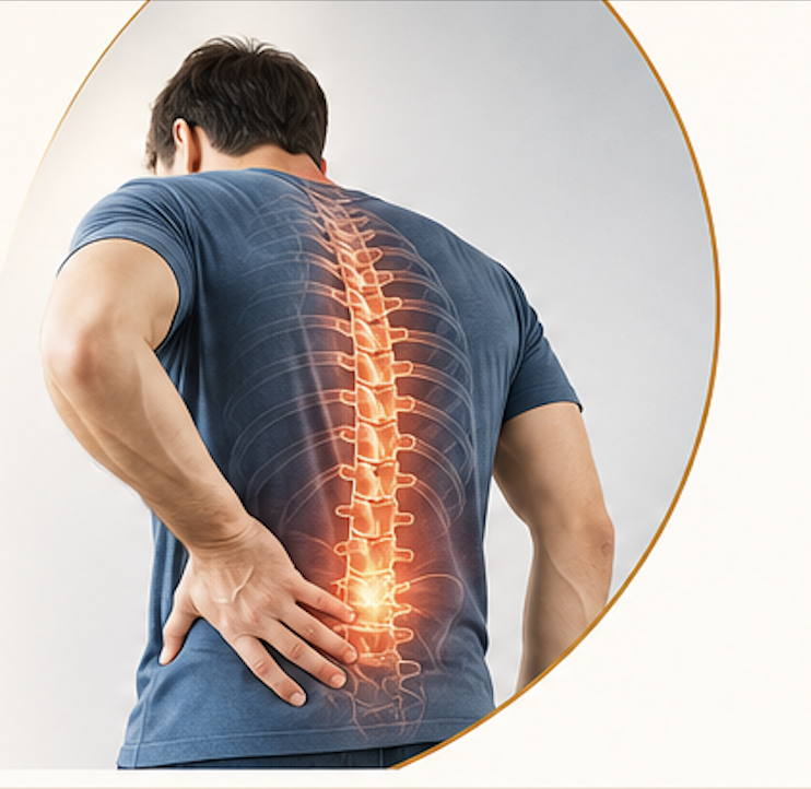
Knee pain has a lifetime prevalence rate of ~45%,1 and represents a source of significant disability and reduced quality of life.
Risk factors for the development of knee pain include
a history of prior injury or surgery, Obesity, and Advancing age.
The most common cause of chronic knee pain is Osteoarthritis (OA), which is characterized by the progressive loss of articular cartilage, with other etiologies including Rheumatoid arthritis, Trauma, crystal arthropathies, and persistent postsurgical pain.
Available treatments for knee pain vary depending on the etiology and diagnosis, but broadly include [A]- physical therapy, [B]-oral medications,[C]- injections, and [D]- surgery.7 Injections for knee pain consist of several types, and may be directed to the soft tissues of the knee joint or the intra-articular joint space. Intra-articular injections encompass a wide range of medications to include anti-inflammatory corticosteroids, pro-inflammatory prolotherapy and platelet-rich plasma (PRP) solutions, viscosupplements. All intra-articular injections require the presence of an intact joint, and are therefore not applicable following total arthroplasty. Knee surgery is similarly heterogeneous and ranges from minimally invasive arthroscopic procedures to open partial or total arthroplasties. Pain due to severe OA is not reliably responsive to conservative therapies, and chronic pain may persist in over 40% of patients who undergo joint replacement, being characterized as severe in 15% of cases. Delivery of radiofrequency (RF) energy to the knee’s nerve supply is a relatively new intervention that can be safely done in the presence of an artificial joint, and may offer an alternative to surgery or surgical revision.
Radiofrequency ablation (RFA) entails the discrete delivery of thermal energy produced by an alternating current to neural tissue, thereby degrading its ability to conduct pain signals. RFA evolved from a therapy primarily employed to alleviate neuropathic pain to one used today predominantly for mechanical joint pain amidst reports of increased pain stemming from deafferentation and neuroma formation.The advent of cooled radiofrequency ablation (CRFA) and the non- ablative pulsed radiofrequency (PRF) herapy have further broadened the clinical utility of RF for chronic pain states. Accepted targets for RF treatment now include most neural structures to include major nerves and ganglia.The use of RF as a treatment for knee pain was who noted complete eradication of pain with intra-articular PRF in a patient with refractory post-traumatic knee pain. This area of pain medicine has evolved in the past decade,
Neuroanatomy of the knee
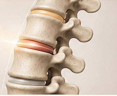
The innervation of the knee joint is complex given that genicular nerves arise from branches of three major nerves: the sciatic, femoral, and obturator, all of which are themselves derived from the lumbar plexus.The sciatic nerve bifurcates into the tibial and common peroneal nerves in the popliteal fossa. The tibial nerve remains in the posterior compartment of the lower leg and gives off the superomedial (SM) and inferomedial (IM) genicular nerves to the posterior aspect of the knee joint. The common peroneal nerve passes into the anterior compartment of the lower leg, and contributes the superior lateral (SL) genicular nerve to the anterior portion of the knee. These genicular branches of the sciatic nerve reliably course in approximation to the periosteum at the medial and lateral junctions of the distal femoral shaft and epicondyles, and at the medial junction of the proximal tibia and epicondyle. The saphenous nerve is a cutaneous sensory branch of the femoral nerve and gives off suprapatellar and infrapatellar (IP) genicular nerves to the anterior portion of the knee. The contribution of the obturator nerve is more variable, but its posterior branch can provide an articular branch to the posterior knee.
Factors associated with radiofrequency ablation treatment outcomes for knee pain and other conditions
| Predictors of success | Predictors of failure |
|---|---|
| Medial compartment osteoarthritis and concordant pain | Greater disease burden (eg, longer duration of symptoms, greater disability) |
| Large and/or multiple lesions | Previous surgery |
| Controlled prognostic blocks | Opioid use |
| Psychopathology | |
| Diffuse pain symptomatology (fibromyalgianess) |
Complications
No major adverse events were reported ,Some patients noted 2–6 weeks of hypoesthesia in the IP region, Injuries to the genicular arterial system are described in the surgical literature, from both open and arthroscopic procedures, and sequelae include pseudoaneurysm formation, hemarthrosis, arteriovenous fistula formation, and patellar osteonecrosis The authors are also aware of several unpublished reports of skin burns, which may arise because of the close proximity of the target nerves to the skin. Other potential complications include those generic to other interventional procedures, namely infection, bleeding, or bruising. Intra-articular approaches also confer the risk of joint sepsis or chondrolysis, but these have not been described following RFA.
Discussion
Chronic knee pain is a recent addition to the growing list of indications for RF, having first been described only a decade ago., the preponderance of evidence suggests that RF denervation can be a safe and effective treatment for chronic knee pain, with the duration of benefit similar to that targeting other joints, ranging between 3 and 12 months.